


Team at University of Aberdeen pioneer new Field Cycling Imager (FCI) scanner able to distinguish tumour material from healthy tissue more accurately than MRI
The treatment of millions of patients could be notably improved thanks to a new scanner developed by scientists at the University of Aberdeen, working in collaboration with NHS Grampian.
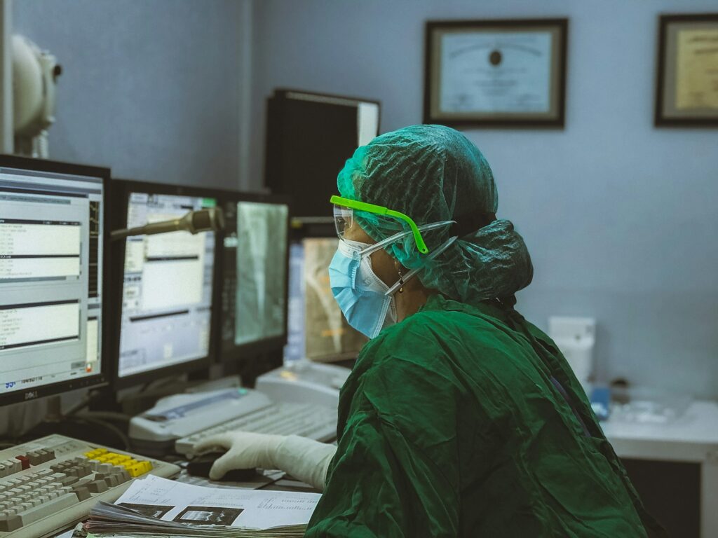
Dr Lionel Broche with the prototype FCI scanner, photo courtesy of University of AberdeenAt the moment, some 15% of women who undergo a lumpectomy must have a second surgery because the edges of the tumour could still be involved. If the new FCI technique can more accurately outline these tumours, few such repeat operations will be needed.
Scanning work of this kind is typically undertaken by magnetic resonance imaging (MRI) scanners, which use magnetic fields, gradients and radio waves to produce images of the organs inside the body. The new FCI system is based on this technology but can vary the strength of the magnetic field being applied during the scan.
This range, including ultra-low magnetic fields, effectively works like using multiple scanners in one. Medical specialists can therefore extract many different types of information about the tissue being scanned and produce a more accurate overall picture.
The University of Aberdeen team tested the FCI system on nine patients with breast cancer, with a total of 10 tumours who were scanned for no more than 60 minutes. The FCI system correctly identified all 10 tumours, but also did better than ultrasound, mammogram and axial MRI scanning in identifying detail, such as ductal carcinoma in situ (DCIS) surrounding the more invasive core.
One major potential benefit of the new technique is that the FCI scanner works without the need to inject ‘contrast’ – coloured dye – into the body of the person to be scanned. That’s good because in some cases such dyes have been associated with allergic reactions and even kidney damage.
The prototype FCI scanner was also shown to be effective at identifying brain damage in patients who have suffered a stroke.
Dr Lionel Broche, Senior Research Fellow in Biomedical Physics at the University of Aberdeen, says: ‘We found that images generated from FCI can characterise breast tumours more accurately. This means it could improve the treatment plan for the patients by improving the accuracy of biopsy procedures by better detecting the type and location of tumours, and by reducing repeated surgery so really, the potential impact of this on patients is extraordinary.
‘My colleagues in the University of Aberdeen built the world’s first clinical MRI in the 1970s so it is both fitting and exciting that we are making waves again with an entirely new type of MRI called Fast Cycling MRI – FCI. This is a truly exciting innovation and as we keep improving the technology for FCI, the potential for clinical applications is limitless.’
Dr Gerald Lip, Consultant Radiologist at NHS Grampian and President of the British Society of Breast Radiology, adds: ‘This data is very promising, and we still need more prospective work, but these results will really support future clinical applications. We treat between 400 and 500 women with breast cancer in NHS Grampian every year and the potential this technology has to reduce the need for women to return for extra surgery is huge, benefitting them and reducing wait times and operating theatre resource. We hope it will have a future role in supporting cancer diagnosis and management.’
In related news:
Leave a Reply